Wrist Tendinitis

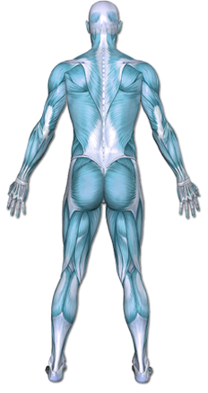
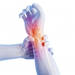
The wrist joint is made of 4 of the small carpal wrist bones where they meet the forearm bones called the radius and the ulna. The wrist moves in multiple planes of motion: up, down, left, right and circles (called circumduction). In order for the wrist to have so much range of motion, there are numerous tendons that cross it. The muscles are in the forearm but the tendons cross the wrist joint so that they can cause the hand to move. With repetitive motion, the tendons can rub on the tissues that create their gliding tunnel. Also, when a wrist has become arthritic the tendons may rub against bony spurs or gliding surfaces that are no longer smooth. Irritation of the tendon can lead to inflammation at the tendon and eventual tendinitis.
The result of overuse, rubbing or inflammation at the wrist is an acute injury and is commonly called tendinitis. Tendinitis refers to the inflammation of the tendon that will usually last for 1-2 weeks after injury. If it doesn’t respond to conservative treatment within two weeks, scar tissue can begin to form within the tendon, leading to the condition of tendinosis. Tendinosis is an advanced state of injury and may lead to having surgical repair of the tendon. Acute tendinitis responds well to anti-inflammatory methods such as ice and rest but tendinosis does not.
This content was created using EBSCO’s Health Library
Tendinopathy is caused by overuse of a muscle-tendon unit. The strain on the tendon causes very tiny tears that accumulate over time. There can also be inflammation. These tears cause pain and can eventually change the structure of the tendon.
This content was created using EBSCO’s Health Library
Factors that increase your chance of developing wrist tendinitis include
- Adults over 40
- In those with rheumatoid arthritis, gout, and psoriatic arthritis
- Repetitive strain (work or recreational tasks)
- Direct trauma
- Prolonged compression (in a cast or brace)
This content was created using EBSCO’s Health Library
The common signs of tendinitis are.
- pain with motion
- inflammation
- good response to anti-inflammatory method
- decline in functional task tolerance
- pain that shoots up the side of the thumb and arm with any activity that requires the hand to open
- dropping objects
- “toothache-type” pain to the side of the thumb and wrist
This content was created using EBSCO’s Health Library
The doctor will ask about your symptoms and medical history. A physical exam will be done. If your symptoms are severe, your doctor may need some images of the tendon and bone. Imaging tests may include:
- X-ray
- MRI scan
- Ultrasound
This content was created using EBSCO’s Health Library
Wrist tendinitis is a common condition seen by both physicians and therapists. If the tendon is not responding to conservative treatment the physician may recommend a cortisone injection to reduce inflammation. The therapist will evaluate for which daily functional tasks are keeping the injury from healing and will offer modifications of those tasks. Therapy may include other anti-inflammatory techniques like education on use of ice and rest, ultrasound treatment or use of a brace. The primary goal of therapy is to reduce the inflammation before it develops into tendinosis and to facilitate a speedy return to daily self-care, work, and recreational tasks.
This content was created using EBSCO’s Health Library
Prevention of wrist tendinitis involves evaluating stressful positions before they become painful.
- When working at a computer desk, use ergonomic principles to reduce side-to-side motion of the wrist, reduce thumb opening and closing and encourage rest breaks from typing or mousing.
- Before and between work activities, do light stretching of the wrist and forearm to prepare the muscles for work.
- Keep the hands and wrists warm as cold tends to increase the risk of tendon irritation. When the wrist becomes painful, seek attention within the first 2 weeks so that the inflammation can be addressed before it becomes chronic.
This content was created using EBSCO’s Health Library
This content was created using EBSCO’s Health Library
- Ashurst JV, Turco DA, Lieb BE. (2010). Tenosynovitis caused by texting: An emerging disease. J Am Osteopath Assoc;110(5):294-6. Dressendorfer R, Richman S. (2013). DeQuervain’s syndrome. CINAHL Rehabilitation Guide; EBSCO Publishing: Apr 05.
- Howell ER. (2012). Conservative care of DeQuervain’s tenosynovitis/tendinopathy in a warehouse worker and recreational cyclist: A case report. J Can Chiropo Assoc;Jun;56(2):121-7
- Klauser AS, Franz M, Arora R, Feuchtner GM, Gruber J, Schirmer M, Jaschke WR, Gabl MF. (2010). Detection of vascularity in wrist tenosynovitis: Power Doppler ultrasound compared with contrast-enhanced grey-scale ultrasound. Arthritis Res Ther;12(6):R209.
- Kwon BC, Choi SJ, Koh SH, Shin DJ, Baek GH. (2010). Sonographic identification of the intracompartmental septum in deQuervain’s disease. Clinc Orthop Relat Res;468(8):2129-34.
- Patterson SM, Picconatto WJ, Alexander JA, Johnson RL. (2011). Conservative treatment of an acute traumatic extensor carpi ulnaris tendon subluxation in a collegiate basketball player: A case report. J Athl Train; 46(5):574-6.
This content was created using EBSCO’s Health Library